At Portland Eye Clinic, we utilize cutting-edge technology to provide high quality eye care.
iCare Tonometer
This state-of-the-art equipment lets us swiftly and easily test for glaucoma without needing the usual “Puff” test or stinging eye drops. It’s a safe and effective method to check your eye pressure.
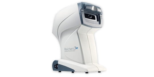
Reichert 7CR Auto Tonometer

The Reichert 7CR from Reichert Technologies uses a patented bi-directional applanation technique to assess the cornea’s biomechanical characteristics, minimizing their effect on IOP measurements. This measurement, known as Corneal Compensated IOP (IOPcc), is less influenced by the cornea’s visco-elastic properties, thickness, or surgeries like LASIK or PRK. Consequently, IOPcc serves as a more reliable indicator of glaucoma risk compared to other tonometry methods, such as Goldmann Applanation.
The Reichert 7CR offers greater clinical accuracy across various patients, including those with normal tension glaucoma, primary open-angle glaucoma, post-LASIK and other refractive surgeries, Fuchs’ dystrophy or corneal edema, keratoconus, and corneas that are thick, thin, or biomechanically unique.
Topcon Fundus Photographer
A fundus, or retinal camera, is a specialized device combining a microscope and a camera to capture images of the central retina, optic disc, and macula. The images produced are high-resolution and magnified, aiding in the detection of eye conditions such as glaucoma, diabetic retinopathy, macular degeneration, optic nerve disease, and more. These images also serve as a baseline for tracking changes in your eye health over time.
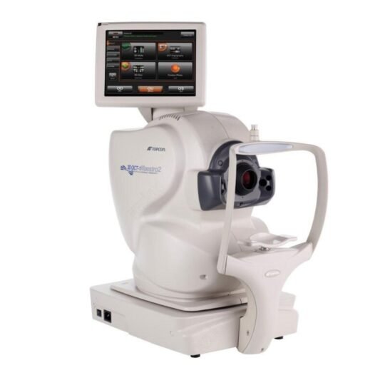
Topcon OCT Maestro
An Optical Coherence Tomography scan, or OCT scan, represents the forefront of imaging technology. Much like ultrasound, it is a diagnostic tool, but it uses light waves instead of sound waves to create highly detailed images of the eye’s structural layers. This scanning laser inspects the layers of the retina and optic nerve for any signs of eye disease in a manner akin to a CT scan of the eye. It uses light and not radiation, making it a key tool for the early detection of conditions like glaucoma, macular degeneration, and diabetic retinal disease. Thanks to an OCT scan, doctors receive color-coded, cross-sectional images of the retina. These precise images are transforming how we detect and treat eye conditions such as wet and dry age-related macular degeneration, glaucoma, retinal detachment, and diabetic retinopathy early on. The OCT scan is a noninvasive and painless procedure that typically takes about 10 minutes to complete in our office. Feel free to contact us to arrange for an OCT scan at your next visit.
At Portland Eye Clinic, we utilize state-of-the-art digital imaging technology to evaluate your eye health. Detecting eye diseases early allows for successful treatment, often preventing significant vision loss. Your retinal images will be stored electronically, providing your eye doctor with a permanent record of your retina’s condition.
Corneal Mapping

Corneal topography, also called photokeratoscopy or videokeratography, is a non-invasive imaging technique used to map the cornea’s surface curvature, which is the eye’s outer layer. Since the cornea accounts for about 70% of the eye’s refractive power, its topography is crucial for assessing vision quality. This three-dimensional map is a valuable tool for ophthalmologists or optometrists, aiding in diagnosing and treating various conditions, planning refractive surgeries like LASIK, evaluating surgical outcomes, and fitting contact lenses accurately. Unlike keratometry, which measures just four points a few millimeters apart, corneal topography evaluates thousands of points across the entire cornea. The procedure is quick and painless.
Corneal Topographer
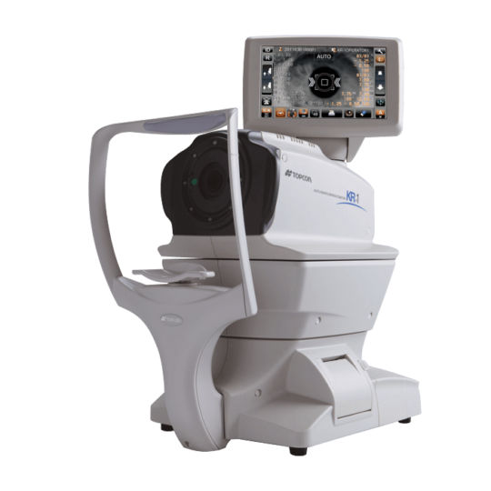
The Topcon Topographer provides precise mapping of the corneal curvature using a computerized Video-Keratometer with Placido rings. This technology is essential for contact lens fitting, refractive surgery, orthokeratology, and overall corneal assessment.
The Medmont E300 Corneal Topographer offers exceptional accuracy and advanced features for doctors. It employs a computerized Video-Keratometer with Placido rings to map the corneal surface. The results from your contact lens exam are crucial for fitting lenses, planning refractive surgeries, and evaluating the cornea’s general health.
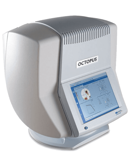
Octopus 600 PRO Computer Perimeter – Visual Field Testing

The Haag-Streit Octopus 600 Pro Computer Perimeter is a versatile and tailored tool for identifying and evaluating visual field defects and managing a range of eye conditions. Its cutting-edge technology and intuitive interface deliver outstanding precision and accuracy in diagnosing and treating ocular issues.
A visual field test checks the range of your peripheral vision to determine if you have any blind spots, peripheral vision loss, or visual field abnormalities. This test is simple and painless, requiring no eye drops—just your ability to follow instructions.
During an initial screening, an optometrist might ask you to focus on a central object, cover one eye, and describe what you see at the edges of your vision. For a detailed examination, special equipment is used. You’ll place your chin on a rest and look straight ahead as lights flash. Your task is to press a button whenever you see a light, which varies in brightness throughout the test. Some flashes are to ensure you’re paying attention. Each eye is tested one at a time, and the process lasts between 15 and 45 minutes. This equipment can generate a computerized map of your visual field, highlighting any areas of concern.


